1 in 3 Adults Has Fatty Liver Disease - The Fix
by Mary Ferrari
“A growing body of research highlights the critical role of intestinal homeostasis in the development and progression of MAFLD.”
SCFAs Regulate Metabolism and Control Inflammation
Metabolic dysfunction-associated fatty liver disease (MAFLD) is now one of the most common chronic liver diseases worldwide, affecting approximately 32% of adults. The disease begins with excess fat accumulating in the liver but can progress to inflammation, liver cell damage, fibrosis, cirrhosis, and even liver cancer. Although genetics, obesity, insulin resistance, and environmental factors contribute to its development, research increasingly points to the gut microbiome as a fundamental part of the disease process. A growing body of research highlights the critical role of intestinal homeostasis in the development and progression of MAFLD. The connection between the gut and liver, known as the gut-liver axis, allows intestinal bacteria and their metabolic products to influence liver function through the portal vein, bile ducts, and bloodstream, this indicates that changes in the intestinal environment can directly affect liver metabolism, inflammation, and overall health.
A healthy gut microbiome helps maintain the intestinal barrier, regulate immune responses, and produce beneficial substances that support metabolic health but microbial dysbiosis can weaken the intestinal barrier by reducing the proteins that hold intestinal cells together. This allows bacterial components and other substances to pass into the bloodstream and travel to the liver. One important example is lipopolysaccharide (LPS), a component of Gram-negative bacteria. LPS activates the TLR4 inflammatory pathway, increasing the production of inflammatory chemicals such as TNF-alpha and IL-6. This creates ongoing inflammation that can damage liver cells and worsen metabolic dysfunction. Studies have also identified changes in bacterial populations in people with MAFLD, including increased Bacteroidetes and decreased Firmicutes.
Short-chain fatty acids (SCFAs), particularly butyrate, are important products of dietary fiber fermentation by gut bacteria and these substances help regulate metabolism and control inflammation. SCFAs inhibit histone deacetylase (HDAC) and activate G-protein-coupled receptors, including GPR43 and GPR109A. These mechanisms promote the differentiation of regulatory T cells (Treg) and the production of IgA antibodies, thereby suppressing inflammation and strengthening the mucosal barrier. Butyrate also helps strengthen the intestinal barrier, improve insulin sensitivity, and reduce fat production in the liver. When dysbiosis reduces SCFA production, the loss of these protective effects can contribute to inflammation and worsening liver disease.
Bile acids provide another important connection between gut bacteria and liver health. Intestinal microbes modify primary bile acids into secondary bile acids, which influence metabolism and immune function. Some secondary bile acids, including 3-oxoLCA, can regulate the balance between Tregs and inflammatory Th17 cells through receptors such as the vitamin D receptor (VDR). Other bile acid pathways involve the FXR and TGR5 receptors, which help regulate glucose metabolism, energy balance, inflammation, and intestinal barrier function. These findings demonstrate that microbial activity influences more than digestion; it also helps control important immune and metabolic processes throughout the body.
Researchers are now investigating treatments that target the gut microbiome and its metabolic products. Hyodeoxycholic acid (HDCA) has shown promise in improving liver fat accumulation and metabolic markers while changing intestinal bacterial populations and bile acid metabolism. Another microbial metabolite, 4-hydroxyphenylacetic acid (4-HPAA), has improved fatty liver conditions in experimental models by activating AMPK, a pathway involved in energy regulation and fat metabolism. AMPK (adenosine monophosphate-activated protein kinase) is widely known as the body’s master metabolic switch or energy sensor.
What AMPK Does
Senses Energy: It monitors the ratio of cellular energy (detecting when ATP drops and AMP/ADP rises due to low energy, exercise, or fasting).
Switches Modes: When activated, it turns off energy-consuming and energy-storing pathways (like fat and cholesterol synthesis) and turns on energy-producing pathways (like glucose uptake and fatty acid oxidation).
Promotes Repair: It triggers cellular housekeeping processes like autophagy (recycling damaged parts) and works in balance with growth pathways like mTOR.
How It Is Activated
Natural Triggers: Vigorous exercise, caloric restriction, fasting, and cold exposure.
Interventions: Certain compounds and plant polyphenols can stimulate AMPK activity.
SCFAs activate AMPK through two primary pathways:
Shifting the Energy Ratio: When cells process and metabolize SCFAs, it temporarily alters cellular respiration, leading to a slight increase in the AMP/ATP ratio. AMPK senses this drop in cellular energy and immediately turns “on”.
Receptor Signaling: SCFAs bind to specific receptors on the surfaces of cells and this binding kicks off a signaling cascade that promotes the phosphorylation (activation) of the AMPK enzyme.
Other approaches include prebiotics, probiotics and fecal microbiota transplantation. Together, these findings suggest that restoring intestinal balance and supporting beneficial microbial metabolites may offer important new approaches to preventing and treating MAFLD.
In conclusion butyrate can:
- Inhibit histone deacetylases (HDACs).
- Activate GPR43 and GPR109A receptors.
- Support regulatory T cells (Tregs) and IgA production.
- Strengthen the intestinal barrier and help reduce inflammation.
Sodium Butyrate
Stopping butyrate supplementation causes intestinal and systemic levels of this short-chain fatty acid to drop back to baseline within a day or two, as your body does not store it long-term.
If you were formula-fed rather than breastfed as an infant, your gut microbiome likely harbors a different baseline community of bacteria, such as lower levels of specific Bifidobacterium species that naturally produce high amounts of butyrate from dietary fiber. Without ongoing supplementation or high-fiber prebiotic foods to feed your native microbes, your gut simply returns to that original microbial and metabolic baseline rather than maintaining elevated butyrate production on its own.
Stopping butyrate supplementation returns your gut to its natural baseline. However, early-life nutrition creates lasting immunological programming. Human milk oligosaccharides (HMOs) promote the long-term development and differentiation of regulatory T cells (Tregs), establishing immune tolerance and dampening chronic inflammation well into adulthood, even without ongoing supplementation.
Dendritic cells act as the ultimate lock and key to the immune system, when specialized compounds like milk oligosaccharides directly interact with human dendritic cells, they turn the key toward long lasting immune tolerance.
Accumulating evidence underlines that milk is a complex signaling and epigenetic imprinting network that promotes stable FoxP3 expression and long-lasting Treg differentiation, crucial postnatal events preventing chronic inflammatory conditions like MAFLD.
Source:
2026
Xue et al. (2026). The Gut-Liver Axis: Microbial Mechanisms and Therapeutic Implications in MAFLD. Frontiers in Cellular and Infection Microbiology, 16:1740163. https://doi.org/10.3389/fcimb.2026.1740163
The gut-liver axis: microbial mechanisms and therapeutic implications in MAFLD
HMO Permanent Shift
- Mechanism: Fucose residue motifs bind specific C-type lectin receptors, primarily DC-SIGN, on mucosal dendritic cells (DCs).
- Outcome: This interaction instructs immature DCs toward a tolerogenic phenotype, upregulating interleukin-10 (IL-10) and transforming growth factor-beta (TGF-beta) secretion, which induces naive CD4+ T cells to differentiate into FoxP3+ Tregs.
- Mechanism: Sialic acid residues bind Siglecs (Sialic acid-binding immunoglobulin-like lectins, particularly Siglec-5 and Siglec-9) expressed on antigen-presenting cells and lymphocytes.
- Outcome: Siglec engagement triggers intracellular immunoreceptor tyrosine-based inhibitory motifs (ITIMs), suppressing pro-inflammatory cascades (such as NF-kB and TNF-alpha) and enhancing immunosuppressive Treg polarization.
- Mechanism: Lacto-N-tetraose backbone configurations serve as ligands for host galectins (e.g., Galectin-1 and Galectin-3).
- Outcome: Galectin activation cross-links cell surface glycans on immune cells, inhibiting effector T-cell activation (Th1/Th17) while promoting Treg survival and expansion.
The Gut Brain Axis Influences Decisions, Intelligence, Cognition and Consciousness
Recent research reveals a fascinating connection between the gut microbiome, neurological function, and conditions like multiple sclerosis and autism. Gut bacteria produce metabolites that enter the bloodstream and influence brain inflammation and neural signaling.
In multiple sclerosis, the immune system attacks myelin the protective insulation surrounding nerve fibers, disrupting electrical signals. Autism involves atypical early brain development and neural connectivity. Scientists are discovering that gut microbes can modulate immune responses and neurotransmitter production, potentially affecting neuroinflammation and brain health across both conditions.
Source:
2026
Dicks LMT, Botha A, Pohl C. Gut bacteria and their metabolites may influence our decisions, intelligence, cognition, and consciousness. Gut Microbes. 2026 Dec 31;18(1):2740906. doi: 10.1080/19490976.2026.2740906. Epub 2026 Oct 3. PMID: 42828728; PMCID: PMC13644589.
Improve immune microenvironment in rheumatoid arthritis via gut microbiota-butyrate-HDAC/NF-κB signaling
Recent research shows that the probiotic Lactobacillus casei alleviates rheumatoid arthritis symptoms by targeting the gut-joint axis. By reshaping the gut microbiome, L. casei boosts production of the short-chain fatty acid butyrate. Butyrate inhibits histone deacetylases (HDACs) and suppresses the NF-κB inflammatory pathway, helping restore immune homeostasis, reduce synovial joint damage, and lower systemic inflammation.
Source:
2026
Ma Y, Li M, Jian L, Peng J, Wen Y, Hao Y, Shu J, Song Y, Li B, Zhang C, Li X, Li G, Wang Y, Peng T, Zhou R. Probiotic Lactobacillus casei improves immune microenvironment in rheumatoid arthritis via gut microbiota-butyrate-HDAC/NF-κB signaling. Gut Microbes. 2026 Dec 31;18(1):2698969. doi: 10.1080/19490976.2026.2698969. Epub 2026 Jul 21. PMID: 42482368; PMCID: PMC13393233.
Butyrate depletion amplifies antibody-driven type I interferon activation in neonatal lupus
Recent research shows that gut microbiome remodeling and butyrate depletion amplify anti-Ro/La antibody-driven type I interferon activation in neonatal lupus erythematosus (NLE). Neonates with NLE exhibit decreased beneficial taxa like Bifidobacterium, altered microbial carbohydrate processing, and reduced systemic butyrate levels. In functional assays, butyrate suppresses anti-Ro/La-associated interferon-α production and downregulates inflammatory proteins linked to cytokine, IL-17, and TNF signaling pathways.
Source:
2026
Sun W, Li Y, Liu X, Yu S, Li W, Wang H, Geng H, Li L, Hu J, Huo J, Zhang W, Fu J, Jin X, Li H, Zhou X, Zhu X. Gut microbiota functional remodeling and butyrate depletion amplify anti-Ro/La antibody-driven type I interferon activation in neonatal lupus. Gut Microbes. 2026 Dec 31;18(1):2728464. doi: 10.1080/19490976.2026.2728464. Epub 2026 Sep 16. PMID: 42747315; PMCID: PMC13596901.
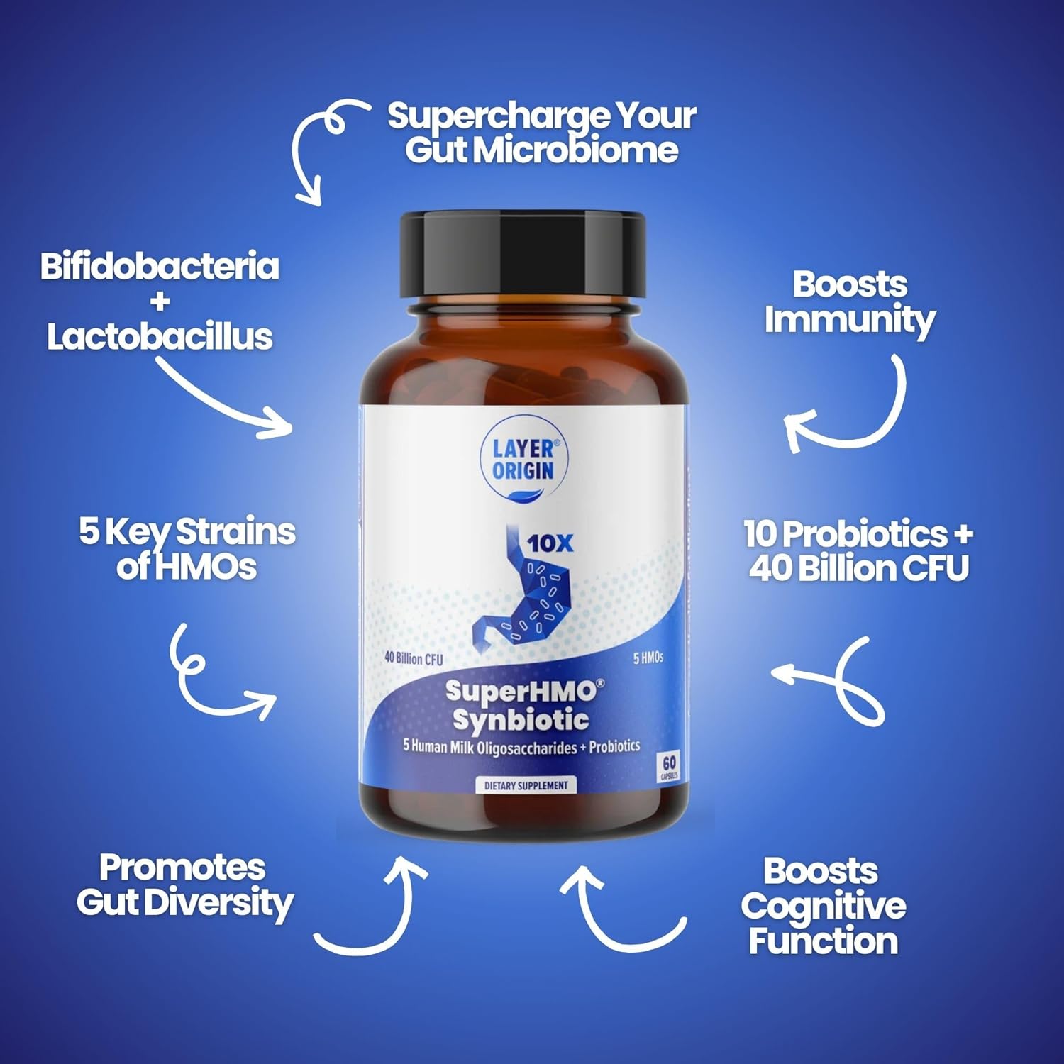
Layer Origins
"Feel better almost immediately I have dysbiosis that has led to SIBO, systemic inflammation, dermatological and joint related issues. On bad flares I get intense joint inflammation and pain to the point of feeling as if I’ve had arthritis for fifty years. Taking this product for just two or three days reverses the inflammation to where I honestly can’t even feel it and my SIBO gas is reduced significantly. This stuff does not make me feel bloated and I can barely tell it is in my shaker" J.